Visit our youtube channel – https://www.youtube.com/@muvis-tomography or see below for examples of our photorealsitic 3D rendering movies.

Cavitation damage on metallic impellor blades
Volume rendering showing cavitation damage on metallic impellor blades
- Sample diameter: ~300 mm
- Scanner: custom 450 kVp/225 kVp hutch
- Imaging mode: CLARITy (scatter-free imaging)
Sample owner: Dr Nicola Symonds – More information

Dissolution behaviour of a 3D printed dosage form captured with time-resolved CT imaging
Time-resolved volume rendering of a 3D printed showing the volumetric changes of a printed dosage form during consequent exposure in media that simulated exposure to gastrointestinal environment.
- Scanner used: Modified 225 kVp Nikon/Xtek HMX
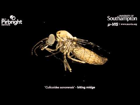
High resolution X-Ray CT imagining of a “Culicoides sonorensis” (a.k.a. biting midge)
- Sample: Culicoides sonorensis
- Staining: Phosphotungstic acid (PTA)
- Sample size: aprox.[1.0 x 1.0 x 0.5] mm
- Scanner: ZEISS Xradia 510 Versa 3D X-ray microscope
Sample owner: Dr Pippa Hawes / Dr. Anthony Wilson (The Pirbright Institute; pirbright.ac.uk)

Inspection of a marine engine injector
- Sample: Marine engine injector
- Sample size: diameter: 20 mm; length 130 mm
- Scanner: custom 450 kVp/225 kVp hutch
Sample owner: Dr. Nicola Symonds, nC2 | Read more about this study here

Mouse embryo at 16 days post fertilisation
µCT imaging, segmentation and reconstruction of a mouse embryo at 16 days post fertilisation.
- Scanner: Nikon Med-X prototype
Data processing by David Chatelet, Biomedical Imaging Unit, University of Southampton
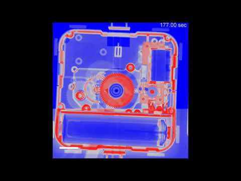
Real-time X-Ray videography of a working clock
- Sample: wall clock
- Sample size: 270 mm (diameter)
- Scanner: custom 450 kVp/225 kVp hutch
- Imaging mode: Flat panel & 225 kV source
Sample owner: μ-VIS
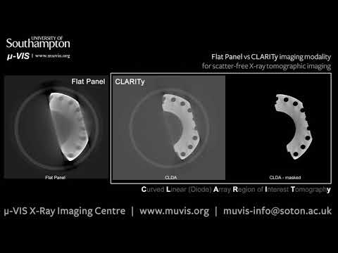
Scatter-free X-ray tomography: introducing CLARITy
CLARITy imaging mode is a single slice -based scatter-free imaging modality that allows stunning image sharpness and contrast of dense specimens
- Scanner: custom 450 kVp/225 kVp Hutch
- Imaging mode: CLARITy (450kV source + CLDA detector)
- Sample: additive manufactured curved stainless steal pipe with flanges;
- Specimen size: 50 mm diameter

Stereoscopic rendering of a Bumblebee
- Sample: Bumblebee
- Resolution: 11.8 μm
- Sample size: aprox. 20mm (length)
- Staining: Non stained
- Scanner: Custom 225 kVp Nikon/Metris HMX ST
Sample owner: Dr. Richard P. Boardman (μ-VIS)
Synchrotron Radiation µ-CT imaging of the planktonic foraminifera Orbulina universa
- Sample: planktonic foraminifera (Orbulina universa)
- Sample diameter: 500 μm
- Sample composition: CaCO3
- Facility: TOMCAT beamline at the SLS at the Paul Scherrer Institute
Sample owner: Dr Thomas H. G. Ezard (University of Southampton)
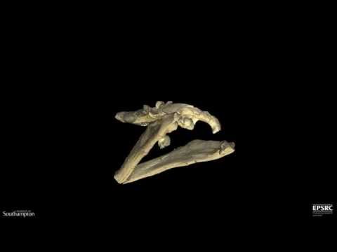
The Weymouth Bay Pliosaur
- Sample: fossilised bone
- Sample size: 2.4m (length)
- Scanner: custom 450 kVp/225 kVp hutch
Sample owner: Dorset County Museum | Read more on BBC and Dorset County Museum websites

Treble recorder made by Peter Bressan
- Sample: Bressan Alto Recorder
- Sample diameter: length: 400 mm; diameter: 20 mm
- Sample composition: wood
- Scanner: Custom 225 kVp; Nikon/Metris HMX ST
Sample owner: Andrew Lamb (Oxford university Bate Collection) | Read more about this project here
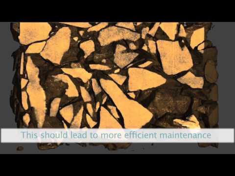
X-CT of a railway ballast for particle analysis
- Sample: Railway ballast (core)
- Voxel size: ~ 300 μm
- Sample size: aprox. 300 mm (length) x 400 mm (diameter)
- Scanner: Custom 450/225 kVp Hutch
Sample owner: Dr. Sharif Ahmed